The Electron Microscopy Facility at the Université de Montréal is open-access facility for the academic and industrial scientific community.

Services

  • Consultation for experimental design

  • Sample preparation

  • Training and instrument access

  • Data analysis

Technics

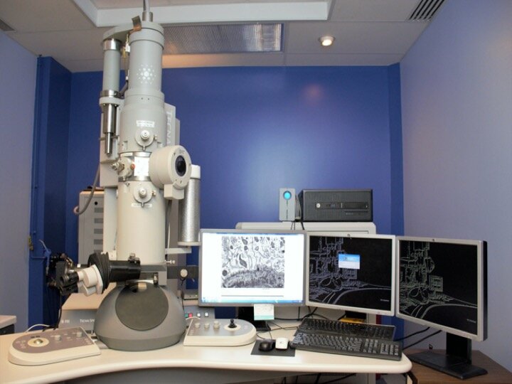
  • Routine and high resolution microscopy

  • Correlative microscopy
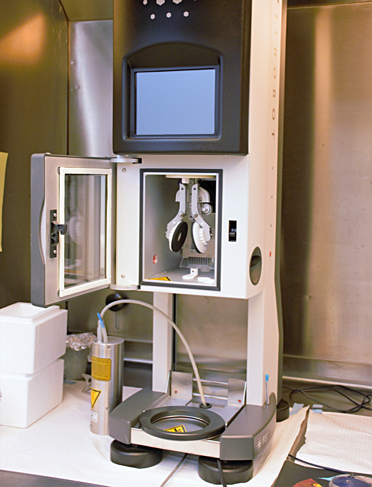
  • Cryo-microscopy

  • Tomography

  • Supramolecular complexes

  • Negative staining

  • Cytochemistry/ Colloidal gold

  • Sample embedding


Acknowledgments

Publication throughput from the facility is important for the funding and sustainability of the Electron Microscopy Facility.

After the paper is published, please complete and send us a PDF copy of the publication or complete citation to Antonio Nanci

Here is a suggested text

o   This work was supported by the Electron Imaging Facility, Faculty of dental medicine, Université de Montréal, Québec, Canada.

o   We thank XX (supporting person’s name) for assistance with imaging/sample preparation/analysisThis work was supported by the Electron Imaging Facility, Faculty of dental medicine, Université de Montréal, Québec, Canada.

EQUIPMENTS

Instrument for imaging

IMG_0684.jpeg

Hitachi Regulus 8220

Ultra-high resolution scanning electron microscope equipped with Oxford energy-dispersive spectroscopy system

Sans titre.jpg

Philips Tecnai 12

Transmission electron microscope equipped with a 4K FEI Eagle camera and 2K AMT camera. Equipped for Cryo-microscopy and tomography

Instruments for sample preparation

cryostat.png

Leica CM1850

Cryostat

microtome.png

Reichert- jung ultramicrotome ultracut E

Semi- and ultra thin sectioning

cpd.png

Leica EM CPD300

Critical point dryer for biological samples

evap.png

Leica EM ACE600

High vacuum sputter coater carbon/platinum/gold

Glow discharge

vitrobot.png

FEI Vitrobot Mark IV

Vitrification of aqueous suspensions

pelco_easiglow_glow_discharge_cleaning_system.jpg
 

Pelco Easiglow

Glow discharge cleaning system


INSTRUMENTS RESERVATION

Access to online reservation system is provided following training on the instrument

INSTRUMENT FEES

Contact us to discuss different payment options for long -term usage or multiple instrument usage.

NOTE: Sample preparation service is temporarily suspended

CONTACT US

Dr Nanci.jpg

Dr Antonio Nanci

Director

Canada Research Chair in Calcified Tissues and Biomaterials, and Structural Imaging

antonio.nanci@umontreal.ca

514-343-5846 

site web

FB_IMG_1495742649013.jpg

Dainelys Guadarrama Bello

Training and access to SEM

dainelys.guadarrama.bello@umontreal.ca

514-343-5231

Ms. Guadarrama is currently on extended leave.

For any inquiries, please contact Ms. Katia Ponce via email at kj.ponce@umontreal.ca

In collaboration with :

Scientific Platforms Biochemistry and Molecular Medicine (BMM), Université de Montréal

Facility for Electron Microscopy Research, McGill University